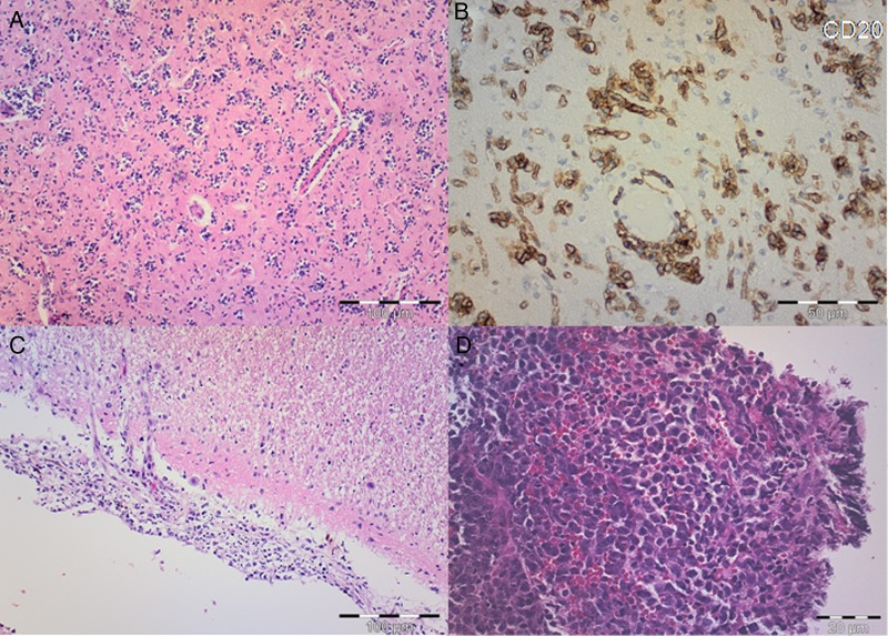
Fig. 2.
(A) Hematoxylin and eosin (H&E) staining shows scattered atypical lymphocytes with diffuse infiltration of the parenchyma and perivascular distribution. Scale bar, 100 µm. (B) On immunohistochemical staining, atypical lymphoid cells are strongly stained with CD20. Scale bar, 50 µm. (C) H&E shows atypical lymphocytes infiltrating meninges. Scale bar, 100 µm. (D) H&E microphotograph of typical nodular primary central nervous system lymphoma with atypical lymphocytes invading brain parenchyma in compact cellular aggregates. Scale bar, 20 µm.

