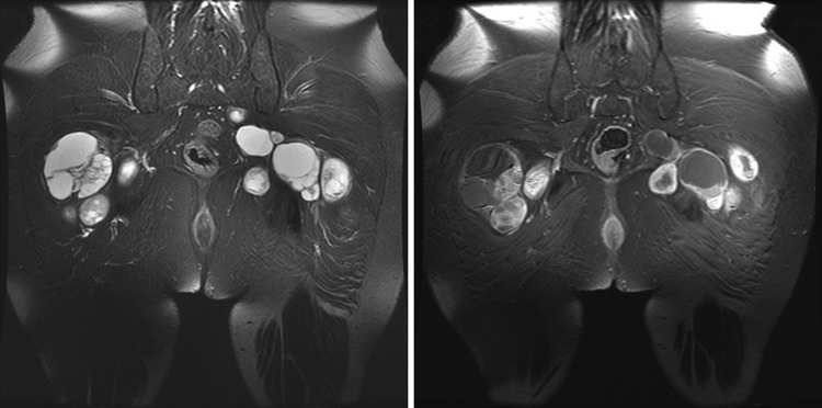
Fig. 3.
Representative MRI scan of the multiple schwannomas in a patient with schwannomatosis. Schwannomas appear hyperintense on T2-weighted images and enhance after administration of gadolinium contrast. The enhancement pattern can range from homogenous to heterogenous. Cysts may develop within some tumors.

