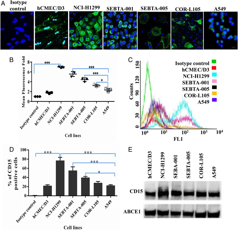
Fig. 1.
Extracellular expression of CD15 in brain endothelial and lung cancer cell lines. (A) Representative immunocytochemical images showing extracellular expression of CD15 in human brain endothelial cells (hCMEC/D3), human non–small cell lung cancer cells (NSCLC) metastatic cells obtained from cervical lymph node (NCI-H1299), brain (SEBTA-001 and SEBTA-005) and in nonmetastatic NSCLC cells (A549 and COR-L105). (B) Semi-quantitative analysis of CD15 expression from confocal images (A) using Zeiss ZEN image software. (C) Representative flow cytometric histogram. (D) Flow cytometric analysis of CD15 expression on hCMEC/D3, NCI-H1299, SEBTA-001, SEBTA-005, A549, and COR-L105. CD15 was highly expressed on NCI-H1299 and SEBTA-001 with less expression on COR-L105 and SEBTA-005, which expressed relatively the same amount. N = 3, ***P < .0001, **P < .001 and *P < .01. There was also less CD15 expression on A549 and hCMEC/D3 cells. (E) Western blot of proteins from the cell lines showed highest CD15 expression in NCI-H1299, followed by SEBTA-001, SEBTA-005, COR-L105, A549, and hCMEC/D3. ABCE1 was used as a protein loading control.

