Fig. 5.
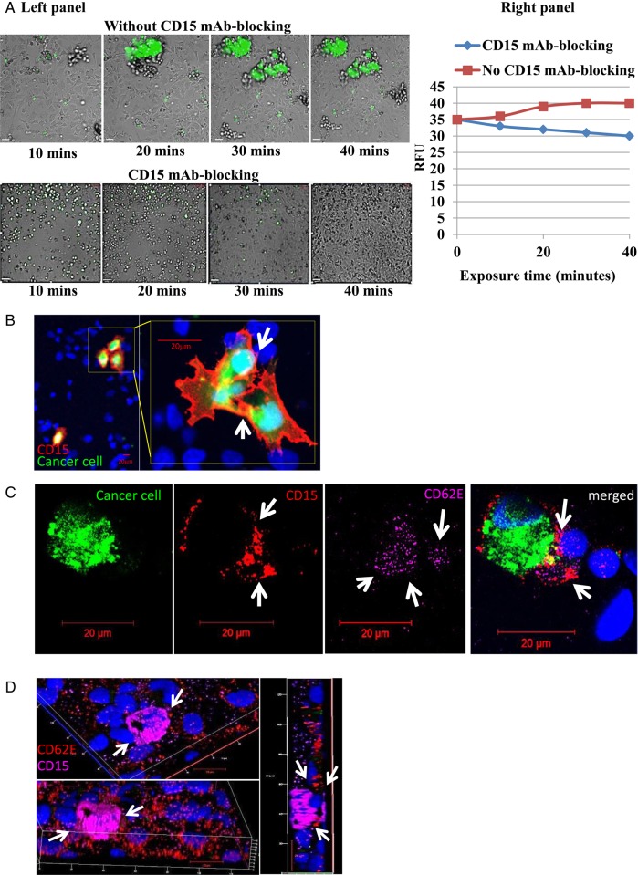
CD15 mAb-blocking reduces adhesion of non–small cell lung cancer (NSCLC) cells under dynamic conditions. (A, left panel) A dynamic cell adhesion assay was carried out on highly metastatic brain cells (SEBTA-001) using an AxioVert 200 M microscope (C. Zeiss) within an environmentally controlled incubator. SEBTA-001 cells were incubated with isotype control (IgM) or CD15 mAb followed by perfusion of 1 × 10⁶ cells over a monolayer of hCMEC/D3 cells at 2.5 dyn/cm2 for 40 minutes. Phase contrast and fluorescent images were acquired at real time every 10 minutes with an X5 objective using Volocity software. Scale bar = 20 μm. (A, right panel) Representation of A, left panel in relative fluorescent units of SEBTA-001 cell adhesion with and without CD15 mAb for 10, 20, 30, and 40 minute time points. (B–D) Confocal images of green fluorescently labeled adherent brain to lung metastatic cancer (SEBT-001) cells cultured on a monolayer of hCMEC/D3 cells (blue). (B) CD15 expression (red) on the edges of SEBT-001 (green). ICC images showed expression of CD15 (red) on the adherent cancer cells (green) on a monolayer of human brain endothelial cells stained with Hoechst blue C: The merged image shows expression of CD15 (red) on adherent tumor cells (SEBTA-001) (green) on an activated monolayer of human brain endothelial cells (blue) expressing CD62E (purple). (D) Optical sections of 3-dimensional confocal image created from z-stack. The top and lower views represent one image at different angles through the z-stack, showing an adherent SEBTA-001 expressing CD15 (purple) on an activated monolayer of brain endothelial cells expressing CD62E (red). Right-side view represents an optical section through the depicted z-stack showing the precise interaction between CD15 and CD62E during NSCLC-brain endothelium cell adhesion.

