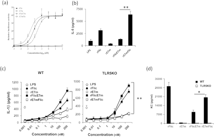
Figure 2. Activation of TLR5 signaling by recombinant proteins in vitro and in vivo.
(a) The relative luciferase activity of cell extracts was analyzed using the dual-luciferase reporter assay system. A total of 1.25 × 10⁵ transfected HEK293/hTLR5 cells/well were stimulated with recombinant proteins at the indicated concentrations for 24 hr. The luciferase activities relative to the control (stimulated with medium alone) were measured. (b) THP-1 cells were treated with 100 nM of recombinant proteins for 24 hr. Supernatants were collected, and the amount of IL-8 was measured by ELISA. (c) WT and TLR5KO BMDCs were seeded at a density of 2 × 10⁵ cells/well. The target protein or LPS (0.1 μg/ml) was added to the LCM medium at the indicated concentration for co-culture with the cells. Cells in medium alone served as the negative control. After 24 hr, the amount of IL-1β in the supernatant was determined. (d) KC levels in the sera were measured by ELISA as described in the “Materials and Methods.” All of the data are expressed as the means ± SEM of three independent tests. * and ** indicate p < 0.05 and 0.01, respectively.

