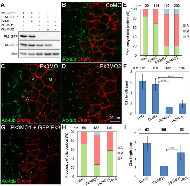
Figure 2. Pk3 is required for the posterior localization and growth of GRP cilia.
(A) Efficiency of the Pk3 knockdown. Embryos were injected with Pk3-GFP RNA (2 ng) that contains MO target sites, control FLAG-GFP RNA (0.1 ng), control MO (CoMO, 80 ng), Pk3MO1 (15 ng), or Pk3MO2 (80 ng) as indicated. Embryo lysates obtained at stage 11 were immunoblotted with anti-GFP antibody. α-tubulin (αtub) is a control for loading. (B–I) Effects of Pk3 depletion (B–F) and rescue (G–I) on GRP cilia position and length. Embryos were injected at the 4–8 cell stage with CoMO (B), Pk3MO1 (C), Pk3MO2 (D) or Pk3MO1 plus GFP-Pk3 RNA (10 pg, G). GRP explants were prepared from stage 17 embryos and stained with anti-acetylated α-tubulin (Ac-tub) antibody to visualize cilia. Coinjection of membrane-associated mCherry RNA (Cherry, 100 pg) marks cell boundaries. (E,H) Percentage of cells with the indicated cilia position. Representative cells with the anterior (A), middle (M) or posterior (P) position of cilia are indicated in (C). Significance was assessed by two-tailed t test comparing the frequencies of cilia positioned posteriorly (green). (E) Pk3MO1 to CoMO: p = 0.0003, Pk3MO2 to CoMO: p < 0.0001. (H) Pk3MO1+Pk3 to Pk3MO1: p = 0.0002. (F,I) Cilia length in GRP cells depleted of Pk3, presented as means +/− s.d. ****p < 0.0001, two-tailed t test. Representative images from three to five experiments are shown, and at least 14 explants were examined per group in each experiment. The effects of Pk3MO1 and Pk3MO2 were evident in approximately 90% of the explants. Co-expression of GFP-Pk3 with Pk3MO1 reduced the frequency of short cilia phenotype to 36% (n = 58). (E,F,H,I) Data were collected from 6 to 10 explants per group in three independent experiments.

