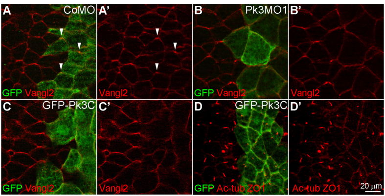
Figure 3. Pk3 is required for the anterior polarization of Vangl2.
(A,B) Immunostaining of Vangl2 (arrowheads) in stage 15 GRP cells from embryos injected with GFP RNA (0.2 ng, lineage tracer) and CoMO (15 ng, A,A’) or Pk3MO1 (15 ng, B,B’). (C,D) Embryos were injected with 2 ng of GFP-Pk3C RNA, and Vangl2 localization (C,C’) and cilia (marked by Ac-tub, D,D’) were visualized in GRP cells at stage 15 (C) and 17 (D) respectively. ZO-1 co-staining reveals cell boundaries. En face staining is shown, anterior is to the top. Representative images from three independent experiments are shown, with 6–10 explants per group.

