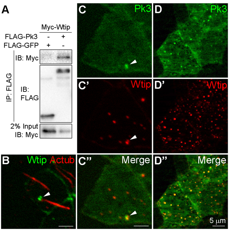
Figure 5. Pk3 and Wtip physically interact and colocalize at the basal body of GRP cells.
(A) Protein interactions between Wtip and Pk3. HEK293T cells expressing Myc-Wtip and FLAG-Pk3 proteins were lysed and immunoprecipitated with anti-FLAG agarose beads. Protein levels were assessed after immunoblotting with anti-FLAG and anti-Myc antibodies. (B) Localization of Wtip at the base of the cilium in GRP cells. Embryos were injected with GFP-Wtip RNA (150 pg). Protein localization is revealed by epifluorescence and staining of acetylated α-tubulin (Ac-tub) in stage 15 GRP explants. (C,D) Colocalization of Pk3 and Wtip in the GRP cells. Embryos were injected with 250 pg of GFP-Pk3 RNA plus 100 pg (C) or 250 pg (D) of HA-RFP-Wtip RNA. Epifluorescence in stage 15 GRP explants is shown.

