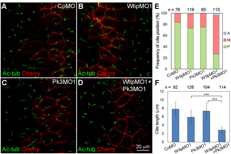
Figure 7. Wtip functionally interacts with Pk3 to regulate GRP cilia growth and positioning.
(A–D) Synergistic effects of Wtip and Pk3 depletion. Embryos injected with membrane-mCherry (Cherry) RNA and CoMO (5 ng, A), WtipMO1 (2.5 ng, B), Pk3MO1 (2.5 ng, C) or WtipMO1 plus Pk3MO1 (D) were stained with anti-acetylated α-tubulin (Ac-tub) antibody to visualize cilia. Membrane-mCherry marks the boundaries of the targeted cells. En face staining is shown, anterior is to the top. Representative images from three independent experiments are shown, with at least 14 explants examined per group in each experiment. While WtipMO1 and Pk3MO1 alone caused short cilia in 20% and none of the explants respectively, co-injection of both MOs increased the frequency of short cilia phenotype to 84% (n = 32). (E) Percentage of cells with the indicated cilia position. The position of each cilium was assigned to the A, M or P location in each cell. Significance was assessed by one-way ANOVA comparing frequencies of posteriorly positioned cilia (green) in the WtipMO1+Pk3MO1 group and the WtipMO1 group (p < 0.001) or the Pk3MO1 group (p < 0.001). (F) Bar graph showing the means +/− s.d. of cilia length in GRP cells injected with MOs. ***p < 0.001, one-way ANOVA. (E,F) Data are collected from six embryos in two independent experiments.

