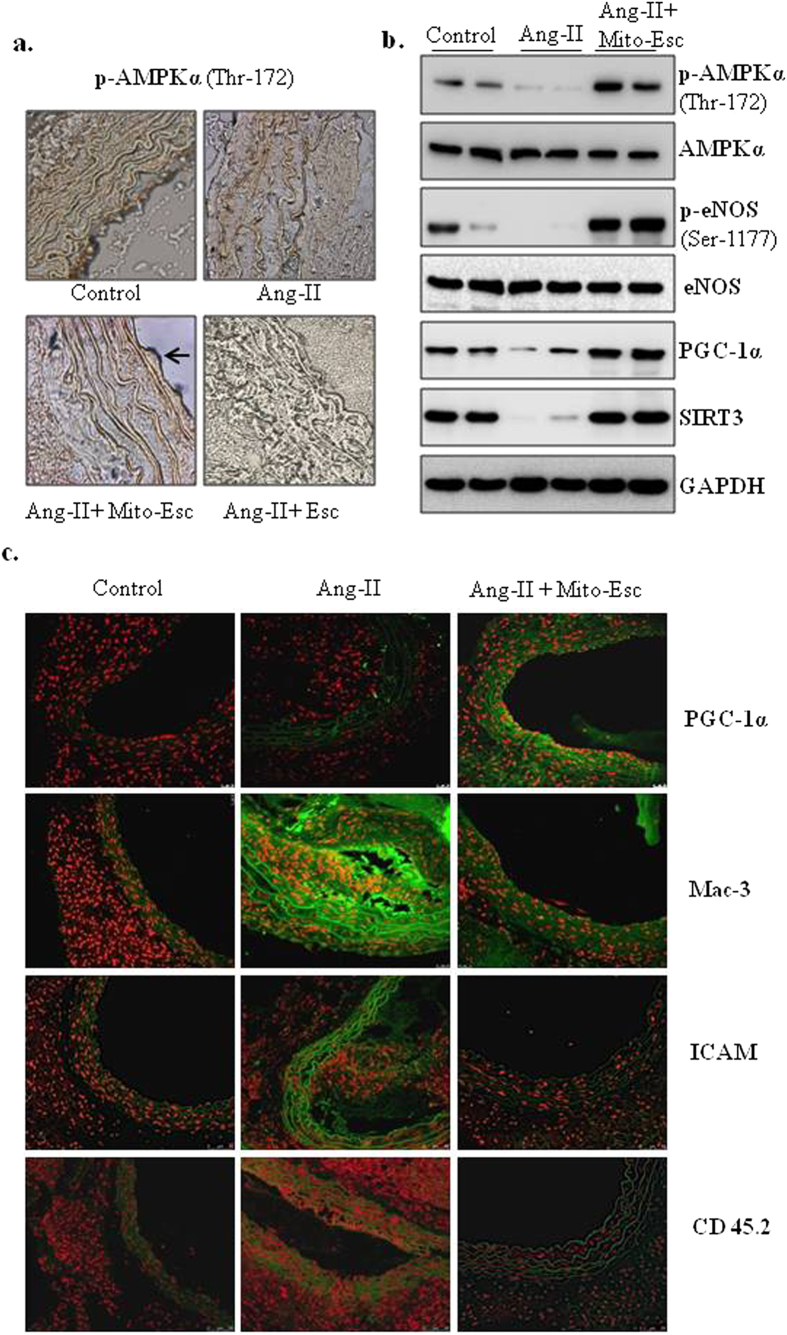
Figure 10. Mito-Esc administration rescues Ang-II-induced alterations in phospho-AMPK, phospho-eNOS, PGC-1α, monocyte infiltration and inflammatory markers in the aorta.
(a) Shows the phospho-AMPKα levels by Immunohistochemistry. (b) Represents phospho-AMPKα, AMPKα, phospho-eNOS, eNOS, PGC-1α and SIRT3 protein levels measured in the aortic tissue homogenate by immunoblotting. Quantification of b is presented in supplementary Fig. S2. (c) Shows the PGC-1α, Mac-3, ICAM and CD45.2 immunofluorescence (green fluorescence represents positive staining as indicated) by confocal microscopy.

