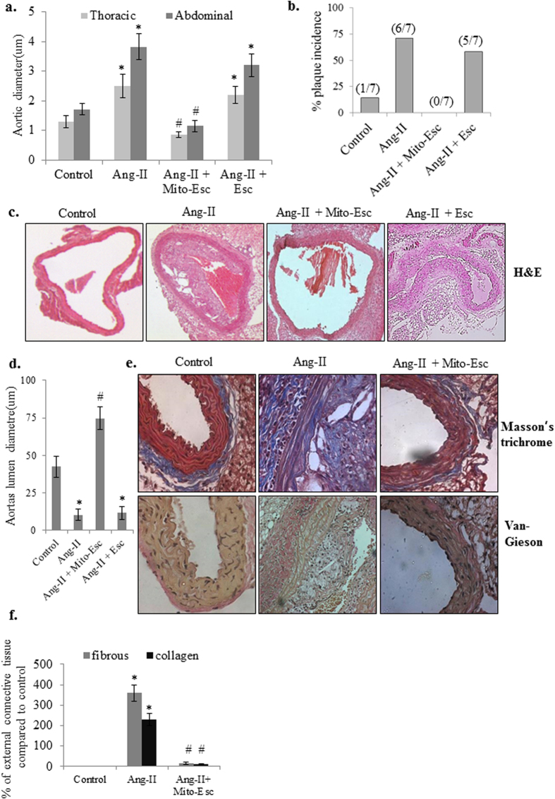
Figure 9. Mito-Esc administration inhibits Ang-II-induced plaque formation in ApoE−/− mice.
(a) Thoracic and abdominal aortic diameters in control, Ang-II, Ang-II + Mito-Esc and Ang-II + Esculetin treated groups. (b) percent plaque incidence. (c) Histopathological images of aorta stained with H&E. (d) Shows aortas lumen diameter. (e) Histopathological images of aortas stained with Masson trichrome and Van Gieson for analyzing fibrous and collagen tissue in the vessel wall. (f) Quantitative analysis of collagen and fibrous tissue in the external region of the vessel wall shown in e. *Significantly different (p < 0.05) compared to control group. #Significantly different (p < 0.05) compared to Ang-II treated group.

