
Figure 1 .
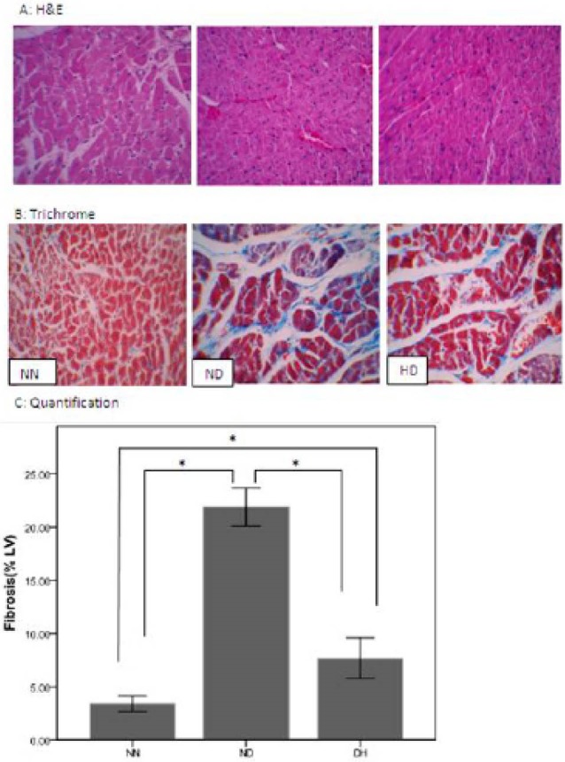
Histopathological analysis of left ventricular (LV) tissue sections stained with (A) Hematoxylin and Eosin (B) Masson trichrome and (C) Quantification of interstitial fibrosis with Masson trichrome stain were measured after 8 week from normoxia normal control group (NN), normoxia diabetes (ND), and chronic intermittent hypoxia diabetes (HD). Where blue = fibrous collagen, and red = cardiomyocytes 400X magnification. *P < 0.05.