
Figure 1.
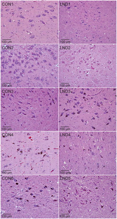
Histopathology in the substantia nigra. Representative hematoxylin and eosin stains of midbrain sections from control (CON1–CON4, CON6) and LND (LND1–LND5) brains are shown. In at least 3 independently stained sections at different levels of the midbrain, there was an impression of reduced neuromelanin pigmentation in 4 cases (LND1, LND2, LND4, LND5), mild or moderate cell loss in 3 cases (LND1, LND4, LND5), and somewhat small soma sizes in 4 cases (LND1, LND2, LND4, LND5).