
Figure 2.
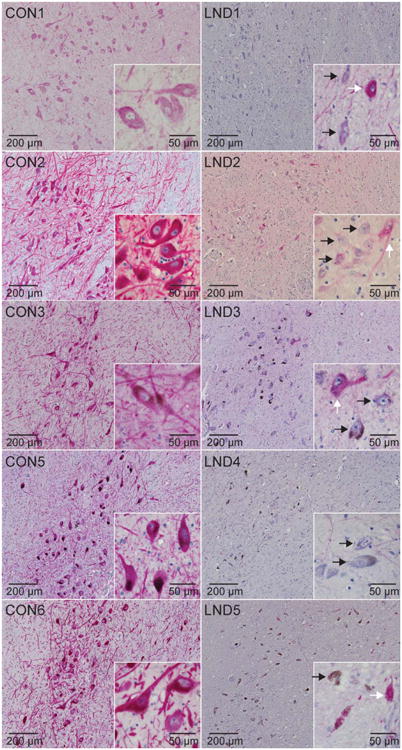
Tyrosine hydroxylase (TH) immunostains of the substantia nigra. Representative TH immunostains of midbrain sections from control (CON1–CON3, CON5, CON6) and LND (LND1–LND5) brains are shown. In at least 3 independently stained sections at different levels of the midbrain, there was an impression that all LND cases showed markedly reduced stain intensity in most pigmented neurons, mixed with a few scattered neurons that stained normally. Melanized neurons with low TH stain intensity (black arrows) can be seen adjacent to neurons with normal staining in the LND cases (white arrows).