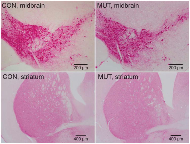
Figure 4.

Tyrosine hydroxylase immunohistochemical stains of the midbrain and striatum in control mice (CON; left column) and hypoxanthine-guanine phosphoribosyltransferase–negative (HGprt−) mice (MUT; right column). Representative sections from the midbrain are shown in the top panels, whereas sections from the striatum are shown in the bottom panels. Consistent results were obtained on tissue from 4 control animals and 4 HGprt− animals.
