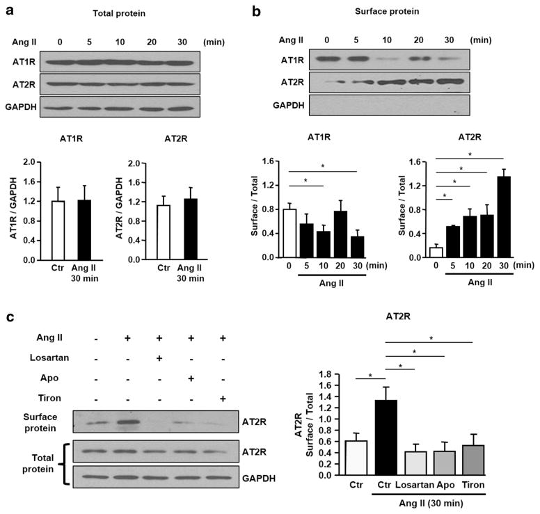
Fig. 3.
Time-dependent expressions of AT1R and AT2R in the surface membrane of LV myocytes following Ang II treatment (3 h) and the effect of ROS scavengers on their expression. a Representative AT1R and AT2R blottings before and after Ang II treatment (5, 10, 20 and 30 min) in LV myocytes. GAPDH was used as a loading control. b Immunoblottings and the mean ratios of AT1R and AT2R (surface/total) at 0, 10, 20 and 30 min after Ang II treatment. c Immunoblotting (left) and the mean ratio of AT2R (surface/total) (right). Losartan (1 μM), apocynin (100 μM) and tiron (1 mM) were pre-treated in LV myocytes, respectively

