Fig. 3.

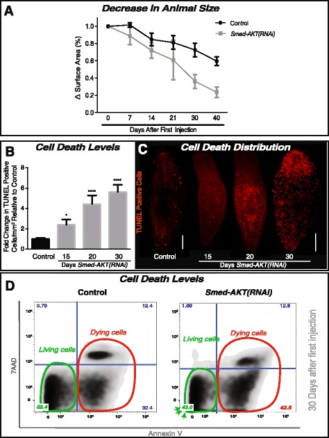
The impairment of Akt leads to increased cell death in S. mediterranea. a The reduction in animal size over time was recorded as the difference in surface area (whole animal) expressed in percentage. Surface area measurements were followed for over 40 days of starvation in both the control and Smed-AKT (RNAi) animal groups. Graph represent the mean ± s.e.m of five independent time courses with ten or more animals per experiment. b Quantification of TUNEL-positive nuclei reveals a gradual increase in cell death from day 15 (~2.5 fold) to 30 days (~6 fold) post first injection relative to its individual control. Graph represent mean ± s.e.m of three biological replicates consisting of five or more animals per experiment. P-value * <0.05 and **** <0.0001, by one way-ANOVA. c Whole-mount immunostaining labeling TUNEL-positive nuclei (cell death) with control on the left and Smed-AKT(RNAi) days 15, 20 and 30 on the right. Each time point consisted of at least two biological replicates with 5 or more animals each. Scale bar 200μm. d Flow cytometry analysis of Annexin V and 7 AAD expression, reveals the frequency distribution between living and dead cells. Annexin V-7 AAD quadrant indicates live cells (green circle). Annexin V + 7 AAD and Annexin V + 7 AAD+ indicates cells that are in early and late stages of cell death, respectively (red circle). The numbers in each quadrant indicate the percentage of cells with that staining profile. All experiments were independently repeated three times with at least ten animals each time
