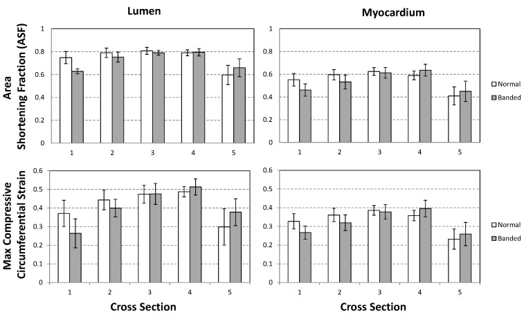
Figure 2.
Comparison of OFT wall motion of control and banded embryos. Analysis was done based on 4D OCT images (control n = 7; banded n = 6), with cross-sectional locations indicated in Figure 1. The data compares ASF on top (Equation (2)) and maximum compressive circumferential strain on bottom (Equation (3)) for the lumen (left) and myocardium (right). Data displays average ± SD.

