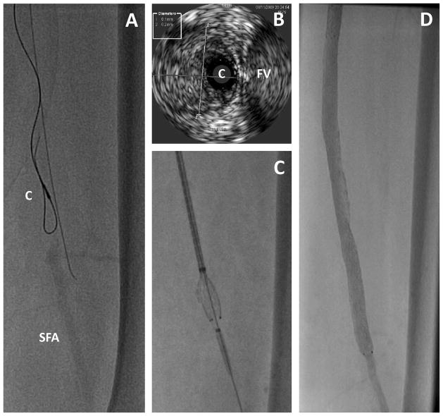
Figure 4.
Intravascular ultrasound used to assess the intra-arterial location while traversing a long occlusion. A. The IVUS catheter (C) is seen over a looped wire in an occluded segment of the mid superficial femoral artery. An adjacent wire is extra-arterial. SFA indicates the distal superficial femoral artery beyond the occlusion. B. IVUS image showing the catheter (C) in the middle of the artery and adjacent to the femoral vein (FV). The diameter of the artery was 6.1 × 6.2 mm, which represents to media and intima and likely overestimates the reference lumen diameter. C. Stent deployment after successfully traversing the occluded artery. D. Final result on angiography.

