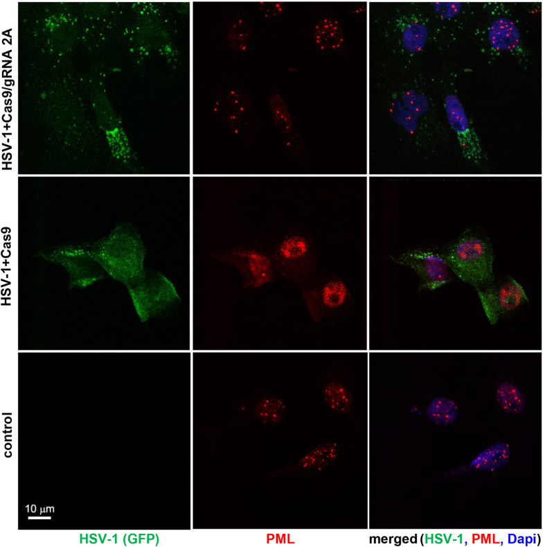
Figure 2. CRISPR/Cas9 by targeting ICP0 expression restores association of PML bodies in L7 cells.
Immunostaining analysis of PML bodies in control, uninfected L7 cells shows the appearance of punctate staining of PML bodies within the nuclei of the cells (lower panel). DAPI staining of nuclei (blue) is shown in the right panels. Infection of Vero L7 cells expressing Cas9 at low MOI with HSV-1/GFP shows complete destruction of the PML and robust replication of HSV-1/GFP as shown in green (middle panels). In L7 cells expressing Cas9/gRNA 2A infection with HSV-1/GFP failed to disrupt formation of punctate PML in the nuclei and significantly reduced the level of viral replication detected in the cells (green, top panels).

