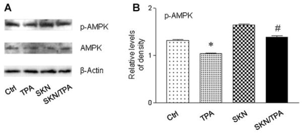
Figure 7.
Shikonin blocked TPA-induced inactivation of AMPK. (A) Western blot analysis to detect AMPK. Cells were treated with TPA (100 nM) or shikonin (1 μM) for 4 h. (B) Quantification of the protein levels of p-AMPK. The level of p-AMPK was normalized to that of β-actin. *P < 0.05 compared with the control (DMSO) treatment; #P < 0.05 compared with the TPA treatment.

