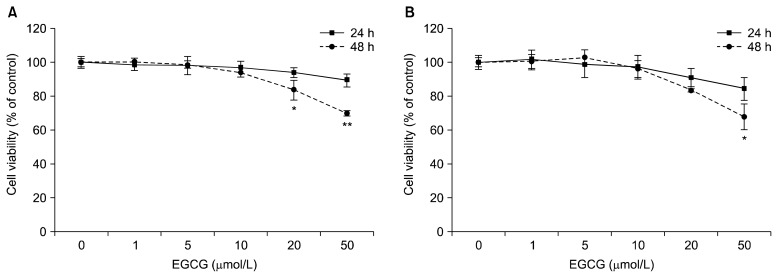
Fig. 1.
Effects of EGCG on cell viability in HepG2 cells (A) and 3T3-L1 adipocytes (B). Cells were treated with 0 (control), 1, 5, 10, 20, or 50 μmol/L of EGCG and incubated for 24 or 48 h. Cell viability was determined using the WST-8 assay. Values are means±SEM (n=3) of three independent experiments. *P<0.05, **P<0.01 vs. control.

