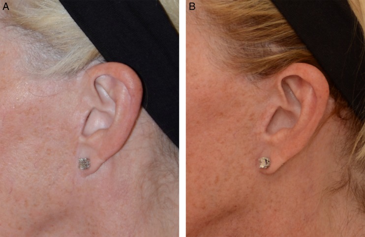
Figure 13.
(A) Preoperative photograph of this 60-year-old woman's ear (same patient in Figure 11B). The scar from a previous facelift (done by another surgeon) is visible and shows a truncated tragus as well as a vertical line over the intertragal incisure. The ear lobe is also attached at the scar. (B) Thirteen-month postoperative photograph after secondary total composite flap facelift. The sideburn has not been raised, and the tragal contours have been re-established. The intertragal incisure has been restored. The ear lobe now hangs normally.

