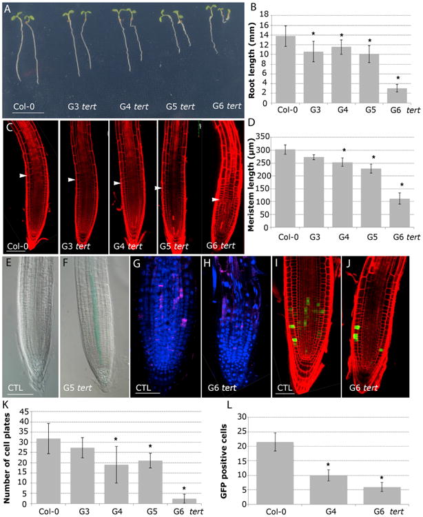
Figure 4. Active Telomerase Is a Requirement to Preserve Cell Division during Root Growth.

(A) Morphology of 6-day-old seedlings of WT and (G3–G6) tert plants. Scale bar, 10 mm. (See also Figure S3.)
(B) Root-length measurements of (G3–G6) tert seedlings compared to the WT. Asterisks denote a statistically significant difference with WT (p < 0.005).
(C) Confocal images of 6-day-old WT and (G3–G6) tert roots stained with PI. Arrows mark the boundary between the proximal meristem and the elongation zone of the root. Scale bars, 100 μm.
(D) The meristem length of 6-day-old roots of (G3–G6) tert mutant generations compared to WT. Asterisks indicate significant differences relative to WT for each day (p < 0.005).
(E and F) GUS staining of 6-day-old roots expressing pICK2/KRP2:GUS (E) and G5 tert; pICK2/KRP2:GUS (F). Scale bar, 50 μm.
(G and H) Whole-mount immunofluorescence using anti-KNOLLE antibodies in WT (G) and G6 tert (H). Scale bar, 50 μm.
(I and J) pCYCB1;1:GFP expression in WT roots (I) and G6 tert (J). Scale bar, 50 μm.
(K) Number of cell plates calculated in whole-mount immunofluorescence with anti-KNOLLE antibodies present in (G3–G6) tert seedlings compared to the WT.
(L) Number of GFP cells in the meristem of pCYCB1;1:GFP (p < 0.001) and G4 tert and G6 tert seedlings (p < 0.001). Asterisks denote a statistically significant difference with the WT (p < 0.001).
