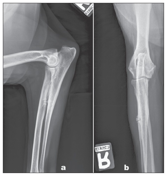
Figure 1.
Lateral (a) and cranio-caudal (b) radiographs of the right antebrachial region of a 10-year-old neutered female golden retriever dog presented with a 2-month history of right forelimb lameness. There was an increase in bone opacity of the radius and ulna density corresponding to the firm swelling that was present, with a suspicion of ulnar proliferation extending towards the radius.

