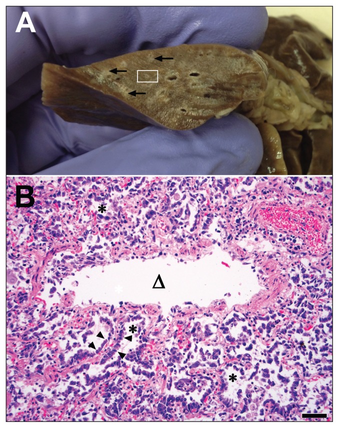
Figure 2.
Gross and microscopic pulmonary lesions in a cat that died from influenza A(H1N1)pdm09 infection. A — Transverse sections of formalin-fixed lung have prominent pale cuffs around bronchioles (arrows). The area enclosed in the white rectangle is magnified in the lower photomicrograph. B — High magnification photomicrograph of a bronchiole (triangle) and adjacent alveoli (asterisks). The bronchiolar epithelium is sloughed and the adjacent alveoli are lined by markedly hyperplastic type II pneumocytes (arrowheads). Bar = 100 μm. Hematoxylin & eosin stain.

