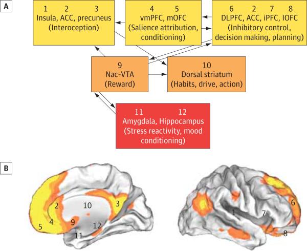
Figure. Schematic Diagram of the Neural Network Necessary for Self-Control and Its Overlap With the Default Mode Network (DMN).
A, The ventromedial prefrontal cortex (vmPFC)/anterior cingulate cortex (ACC), identified by Seo et al as a robust biomarker of alcohol relapse risk, modulates and, in turn, is modulated by the activity of multiple other cortical and subcortical regions whose combined output is used to orchestrate adaptive and flexible goal-directed behaviors. Current evidence suggests that information from cortical and subcortical structures converges toward a single common value representation before passing on to the choice-related motor control circuitry. Modulatory inputs play a critical role in establishing this final common representation with those inputs carrying signals related to interoception, salience attribution, conditioning, executive control, reward, habituation, and stress and mood reactivity. B, In addition, each region has been numbered and its general location tagged onto maps of the human brain (internal and external views) overlaid with orange and yellow pseudocolors that represent the resting state connectivity of the DMN. Regions (vmPFC/ACC and precuneus) that were hyperactive during the neutral conditions and that predicted relapse show significant overlap with the DMN. DLPFC indicates dorsolateral prefrontal cortex; iPFC, inferior prefrontal cortex; lOFC, lateral orbitofrontal cortex; mOFC, medial orbitofrontal cortex; Nac, nucleus accumbens; VTA, ventral tegmental area.

