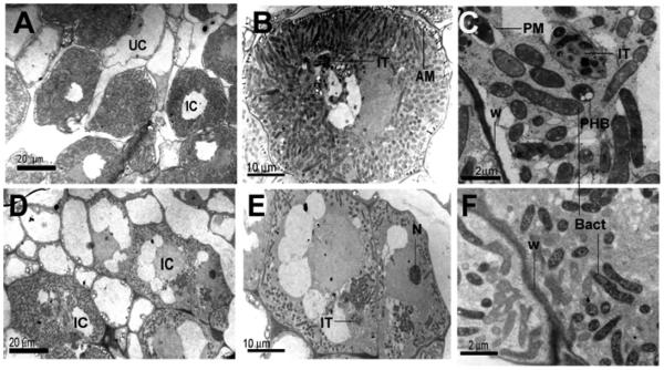
Fig. 8.
Electron micrographs of Medicago truncatula cells from 3-week-old nodules infected by A, B, and C, Rm1021 or D, E, and F, the 1021Δhfq mutant. Nodules were harvested from main roots between the position of the root tip at the time of inoculation (RT1) and the position of the root tip 24 h after inoculation (RT2). IC, Infected cells; UC, uninfected cells; IT, infection threads; W, plant cell wall; N, nucleolus; Bact, bacteroids; AM, amyloplasts; PHB, polyhydroxybutyrate granules. Brightness and contrast of images were adjusted using default settings of Adobe Photoshop version 8.0.

