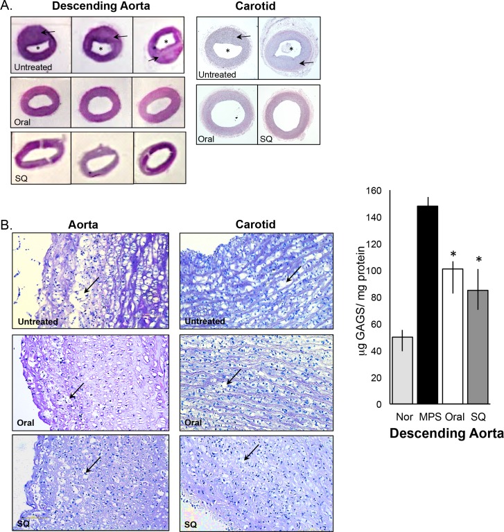
Fig 4. Analysis of the descending aorta and carotid in untreated and PPS-treated MPS I dogs.
Representative images (H&E staining) are shown for untreated, oral, and subQ PPS-treated MPS I dogs (17 months with daily oral treatment and 12 months with biweekly subQ). (A) The intima media of both the descending aorta and carotid is thickened (arrows) in the untreated MPS I dog due to lysosomal storage, resulting in narrowing of the lumen (*) (magnification 1X). PPS treatment decreased the thickness of the intimal media and increased the diameter of the lumen with both modes of administration, but was more significant with subQ treatment (see Tables 2 and 3) (B) Higher magnification (10X) of the descending aorta and carotid artery in untreated and PPS-treated MPS I dogs revealed reduced vacuolization/storage in the treated animals when compared to untreated (arrows). (C) Total GAGs were determined in the descending aorta of normal, untreated and PPS-treated MPS I dogs. PPS treatment significantly reduced GAG storage with both modes of treatment, consistent with the histological evidence of storage reduction. Black columns, untreated MPS I dogs; white columns, oral PPS-treated MPS I dogs; grey columns, subQ PPS-treated MPS I dogs. The vertical lines in each column indicate the ranges. * p = 0.0041

