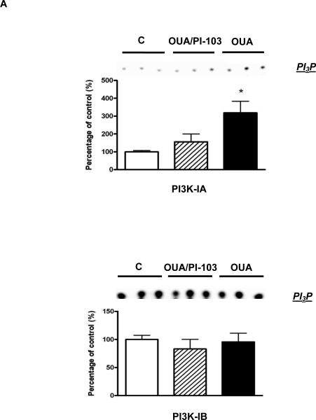
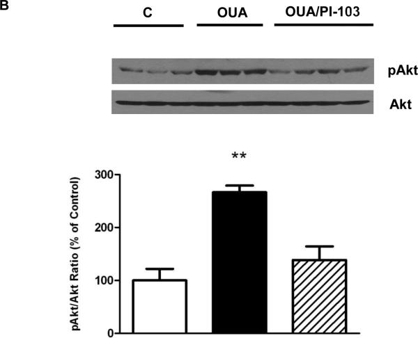
Figure 2. OPC-induced activation of the PI3K-IA/Akt Pathway.
Mouse hearts were perfused with ouabain 10μM for 4 min in the presence or absence of PI-103 100 nM and frozen in liquid nitrogen (Protocol A in figure 1). 2A. PI3K-IA&IB Activity was measured as described in Methods. 2B. Akt. Total Akt and Ser473 phosphorylated Akt (pAkt) contents were evaluated. Values are mean ± SEM (n=3-4). * P<0.05 and ** P < 0.01 vs. control.


