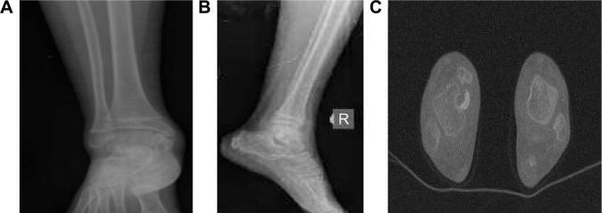
Figure 1.
Ankle images of the patient.
Notes: Localized, irregular osseous mass at the level of anteromedial talus and medial malleolus on AP and lateral ankle radiographs (A and B). Lobulated osteocartilaginous mass appearance in the vicinity of the medial malleolus at the medial aspect of the distal tibia, right side on the axial section of CT (C).
Abbreviations: AP, anterior–posterior; CT, computerized tomography.

