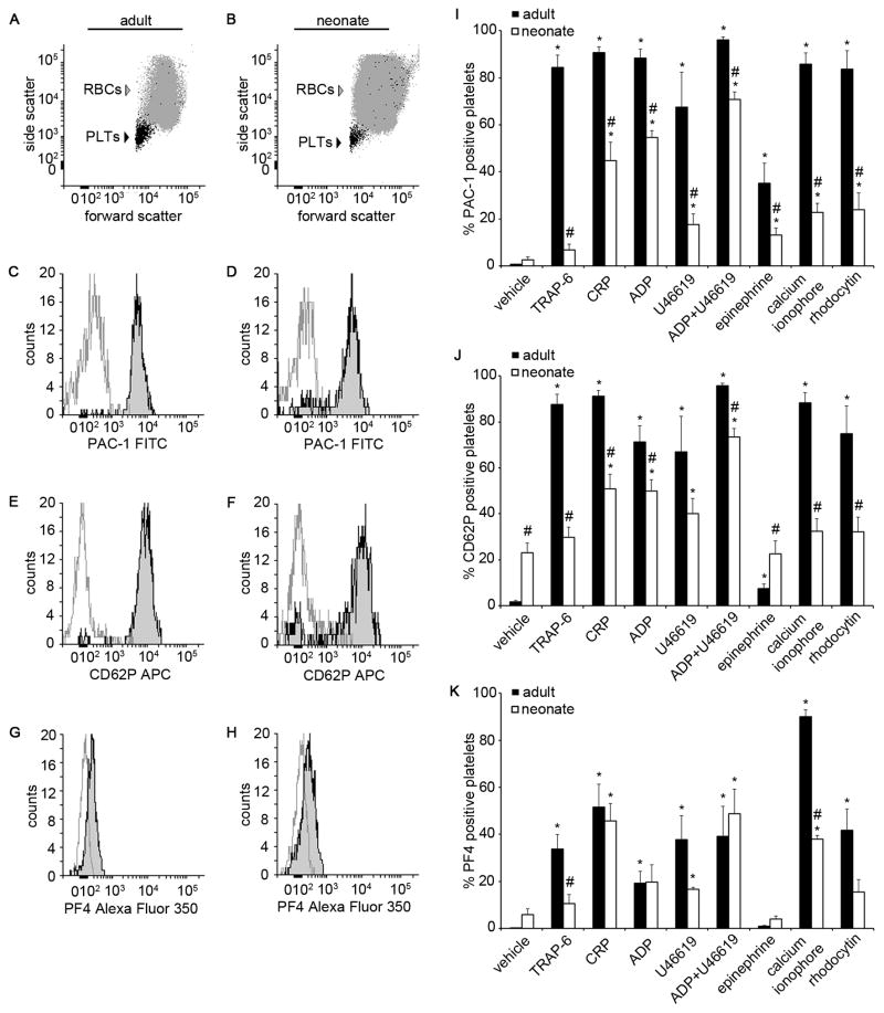
Figure 1. Adult and neonatal whole blood platelet activation.
Representative flow cytometry forward and side scatter dot plots of a whole blood sample from an adult (A) and a neonate (B). Representative histograms of PAC-1-FITC (C and D), CD62P-APC (E and F), and platelet factor 4 (PF4)-Alexa Fluor 350 (G and H) fluorescence intensity of adult and neonatal whole blood treated with adenosine 5′-diphosphate (ADP) + U46619 (10 μM; black line) or vehicle (gray line). Percent of platelets positive for PAC-1-FITC (I), CD62P-APC (J), and PF4-Alexa Fluor 350 (K) in response to thrombin receptor activator peptide-6 (TRAP-6; 10 μM), collagen related peptide (CRP; 10 μg/mL), ADP (10 μM), U46619 (10 μM), ADP+U46619 (10 μM), epinephrine (10 μM), calcium ionophore A23187 (10 μM), rhodocytin (300 nM), or vehicle treatment. Data are represented as mean ± SEM; Nadult = 6 and Nneonate = 8; *P < 0.05 with respect to vehicle treated samples; #P < 0.05 with respect to adult samples.

