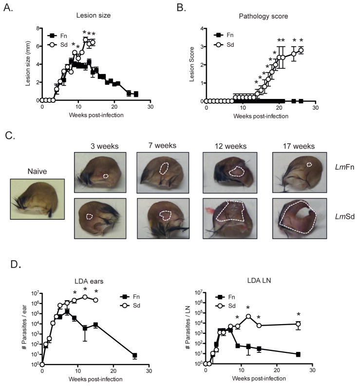
Figure 1. Lm Sd produces a non-healing lesion and severe pathology in C57BL/6 mice.
C57BL/6 mice were infected in the ear dermis with 1000 LmFn or LmSd metacyclic promastigotes. (A) Development of nodular lesions and (B) pathology score (0=no ulceration, 1=ulcer, 2=half ear eroded, 3=ear completely eroded) over the course of infection. (C) Photographs of the ears were taken at different times during the course of infection. Dotted lines circumscribe the nodular lesion. (D) Parasite burden as determined by limiting dilution analysis (LDA) in the ear lesion and dLN over the course of infection. Results shown are the mean ± SD of 10 ears, 5 mice/group, * p<0.05 comparing infection with LmFn and LmSd. The results are representative of at least 3 independent experiments.

