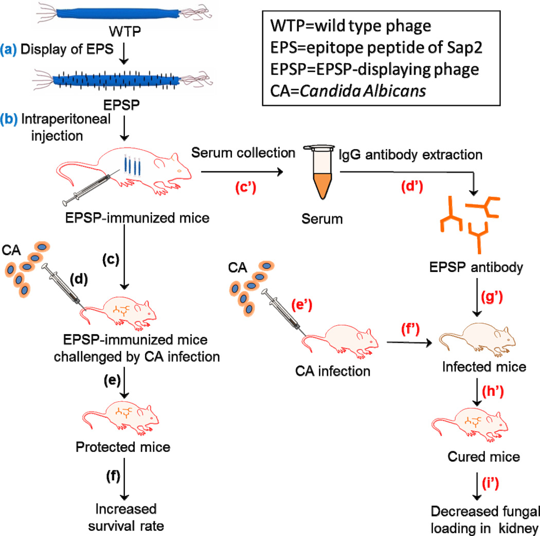
Scheme 1.
Schematic illustration of the general idea using EPSP nanofibers (~900 nm long and ~7 nm wide) as a vaccine for preventing CA infection. Firstly, EPS was displayed on the WTP to form EPSP nanofibers (a), which were intraperitoneally injected into the mice for three times (25 µg/mice each time) to obtain EPSP-immunized mice (b). Then, two approaches were adopted to prove the use of EPSP as a vaccine for preventing CA infection. The first approach is to test if CA cells can infect the EPSP-immunized mice. Briefly, one week after the last mmunization, mice were challenged (c) by injecting 106 CA cells through tail vein (d). It was found that the EPSP-immunized mice were protected from CA infection (e), as evidenced by the decreased fungal loading in kidney, less visceral lession and increased survival rate (f). The second approach is to test if EPSP antibodies collected from the EPSP-immunized mice can be used to cure the CA-infected mice. Briefly, serum was collected from the blood of EPSP-immunized mice (c’), followed by the extraction of IgG from the serum to obtain EPSP antibodies (d’). After the mice were infected by CA cells through tail vein injection (e’ and f’), the EPSP antibodies were injected intravenously into the infected mice (g’), which led to the curing of CA infection (h’) as evidenced by the significantly reduced fungal loading in kidneys (i).

