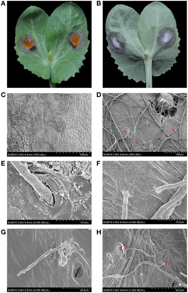
Figure 3.

The infection patterns of A. pinodes ZJ-1 were visualized by scanning electron microscope. (A) The necrotic lesions on the surface of pea leaves (Zhewan-1 cultivar) caused by ZJ-1. (B) The mycelia of ZJ-1 penetrated the leaves and formed velvet on the backside. (C) The light-dark contrasts and physical patterns of boundary between healthy and necrotic tissue. (D) The mycelia patters and penetration structures of ZJ-1 formed on the leaves. (E) The mycelia penetrated the leaves across the stomas. (F,G) The mycelia of ZJ-1formed specific penetration structures and directly pierced leaves. (H) The infective hyphae were able to shuttle back and forth on the leaves and subsequently caused the brownish necrosis and chlorosis symptoms. The voltage and bars were indicated at the bottom of each panel.
