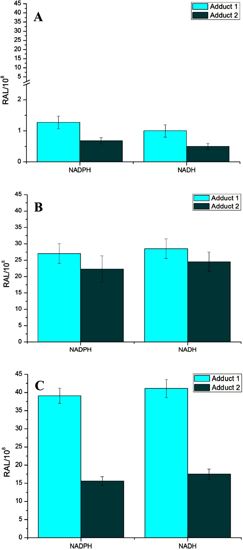
Fig. 4.
DNA adduct formation by BaP activated with microsomes isolated from livers of control rats (a) or rats pretreated with Sudan I (b) or BaP (c) in the presence of either NADPH or NADH. Comparison with previous 32P-postlabeling studies [15, 29] showed that adduct spot 1 has similar chromatographic properties on PEI-cellulose TLC to a guanine adduct derived from reaction with 9-hydroxy-BaP-4,5-oxide [15]. Adduct 2 is dG-N 2-BPDE adduct. For all panels, values represent mean total relative adduct labeling (RAL) ± SD (n = 3; analyses of three independent in vitro incubations)

