. 2016 Jan 28;12(2):188–193. doi: 10.3988/jcn.2016.12.2.188

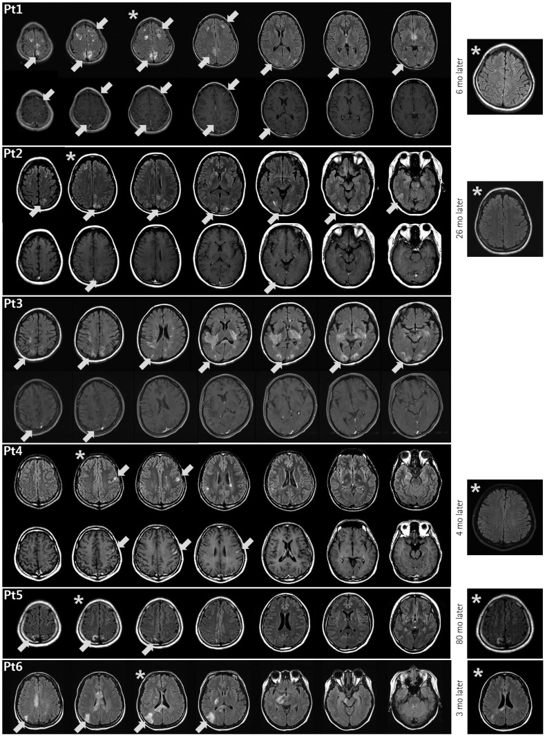
Fig. 1. Brain abnormalities in neuromyelitis optica spectrum disorder (NMOSD) patients on fluid-attenuated inversion recovery (FLAIR) (upper rows) and T1-weighted enhanced (lower rows) magnetic resonance imaging (MRI) scans obtained at the time of cortical lesion presentation. The brain MRI scans of Patient 2 have been described previously.15 The lesions involving the cerebral cortex are accompanied by leptomeningeal (Patients 1, 2, and 3) or cortical (Patient 4) enhancement, or no enhancement (Patients 5 and 6). In addition to the cortex-involving lesions, other characteristic brain lesions of NMOSD are evident. In representative follow-up FLAIR images (Patients 1, 2, 4, 5, and 6), obtained 3 to 80 months after the presentation of the cortex-involving lesions, most of the lesions had disappeared or become faint—compared to the correlated slices in the previous images (*)—in all patients except Patient 5. mo: months.

Fig. 1