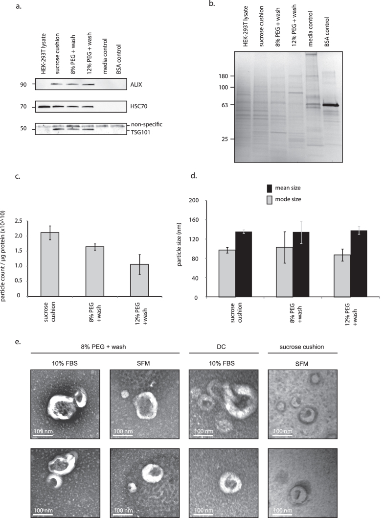
Figure 3. Highly pure extracellular vesicles can be enriched using PEG.
(a) Western blot was used to assess the purity of samples from conditioned serum-free media (SFM). Abundances of exosome markers in PEG-treated samples were comparable to the sucrose cushion isolate. Probing for TSG101 produced a non-specific band at the 55–60 kDa marker; the non-specific band appeared consistently in samples containing only stock medium with 10% FBS, and in the bovine serum albumin (BSA) negative control. (b) Gels were coomassie-stained to compare total protein isolated from sucrose cushion and PEG methods. (c) The 8% PEG + wash method produced highly pure samples, comparable to the sucrose cushion isolates (no statistical difference, p = 0.169). (d) No differences in particle size were observed between treatment groups. (e) Presence of exosome-sized, cup-shaped vesicles was verified by electron microscopy. FBS, fetal bovine serum. DC, differential centrifugation. TEI, Total Exosome Isolation (Life Technologies). All approximate protein masses are represented in kilodaltons (kDa).

