
Fig. 3.
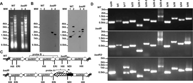
Genetic analyses of badR-deficient B. burgdorferi strains. Total genomic DNA from parental B31-A3 (WT) and badR-deficient strain (badR−) was digested with HindIII (lanes 1 and 3) or EcoRI and NdeI (lanes 2 and 4), separated on a 1% agarose gel, and transferred to nylon membranes (A, B, C). Membranes were hybridized with PCR amplified probes corresponding to the aadA gene (StrR marker) (B) or to a region upstream of the badR gene (C). Schematic representation of the badR region of the chromosome for both WT and badR−. Probes used are indicated with brackets. HIII-HindIII, ERI-EcoRI, ND1-NdeI (D). PCR confirmation to assess plasmid profile was performed on parental and badR-deficient strains. Strains and molecular weight (in base pairs) are indicated on the left (E).