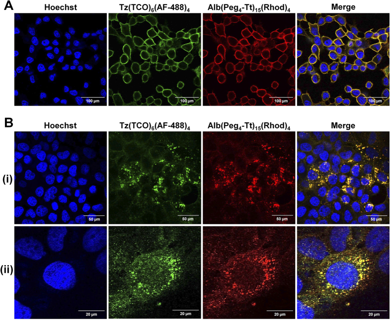
Figure 2. Validation of the internalization strategy by in vitro confocal microscopy.
In vitro confocal fluorescent images of the two-component delivery strategy in HER2(+) BT-474 cells showing (A) cell surface labeling by Tz(TCO)6(AF-488)4 followed by the co-localization of Alb(Peg4-Tt)15(Rhod)4 after 15 min incubation at 20 °C (Scale bar: 100 μm), and (B) rapid cellular internalization after incubation at 37 °C for 4 h (Scale bar: 50 μm). (i) Full field-of-view 63x confocal images, and (ii) a zoomed-in, high-resolution section of the images (Scale bar: 20 μm).

