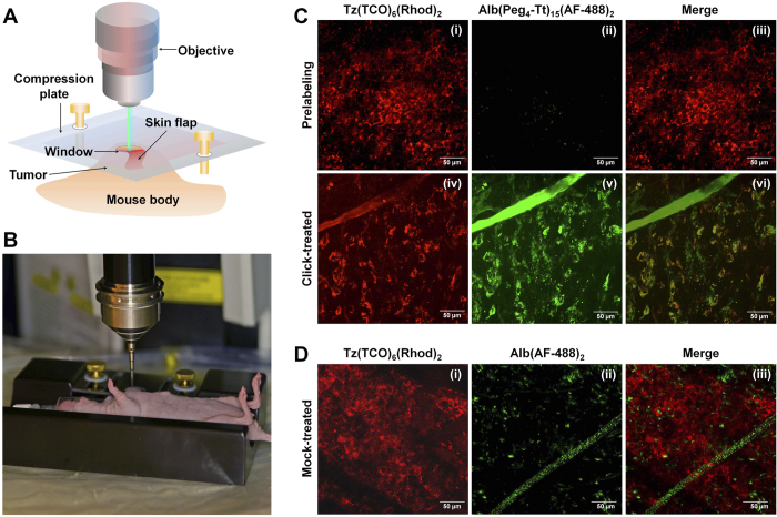
Figure 4. Evaluation of the pre-targeting approach by intravital microscopy.
(A) Schematic view of the intravital microscopic imaging after skin-flap surgery. (B) Custom-made mouse-holder and set-up for intravital microscopy of cancer in mouse models. (C) Intravital multiphoton fluorescence images after the minimally invasive skin-flap surgery in click-treated mice (Scale bar: 50 μm). (i) Tumor uptake of pre-targeting Tz(TCO)6(AF-488)2 in the red channel after 12 h post-injection; (ii) autofluorescence in the green channel; (iii) merging two channels; (iv) tumor uptake of Tz(TCO)6(Rhod)2 after 1.5 h of Alb(Peg4-Tt)15(AF-488)2 injection; (v) tumor uptake of the Alb(Peg4-Tt)15(AF-488)2 delivery component; and (vi) merging of red and green channels shows the co-localization of the two components. (D) Intravital fluorescence images of the control mock therapy. (i) Tumor uptake of pre-targeting Tz(TCO)6(AF-488)2, (ii) control non-reactive Alb(AF-488)2, and (iii) merged images after 1.5 h of Alb(AF-488)2 injection.

