
Figure 1. The evaluation methods of radiation brain necrosis.
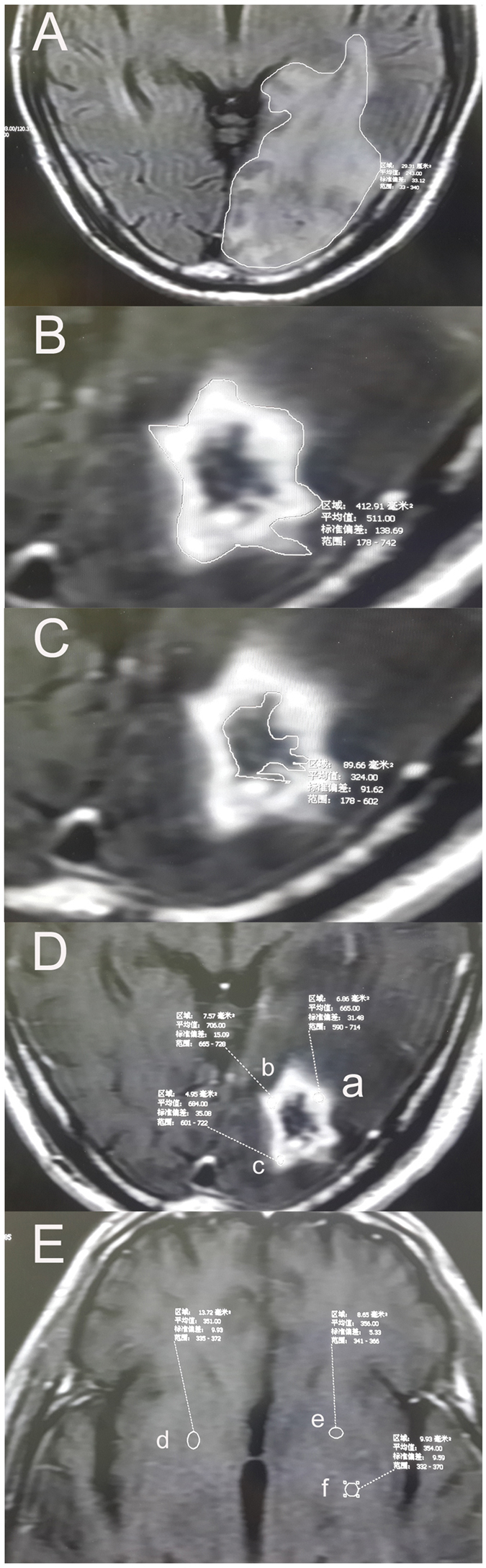
(A) The area of the edema at one layer of the MRI image. (B) The outer edge of radiation necrosis of the brain. (C) The inner edge of radiation necrosis of the brain. (D) Determination of MRI signal in brain necrosis area. (E) Determination of MRI signal in cerebral white matter area at the same level. The volume was calculated using the product of the area of the edema region and the slice thickness. The edema index (EI) was used for evaluation. The EI = Volume A/Volume B. The volume of cerebral necrosis was calculated using the subtraction of the inner diameter volume from the outer diameter volume of the enhancement region of cerebral necrosis(Volume B subtract Volume C). The changes in information in the region of cerebral necrosis used the measurement of the T1 strengthening phase. Three areas in the strengthening region of cerebral necrosis were selected to measure the signal values and to calculate the mean value; in addition, the value was compared with the white matter signal value of the same MRI to eliminate the influences of different strengthening degrees; this ratio was used to measure the changes in signals in the regions of cerebral necrosis before and after treatment.