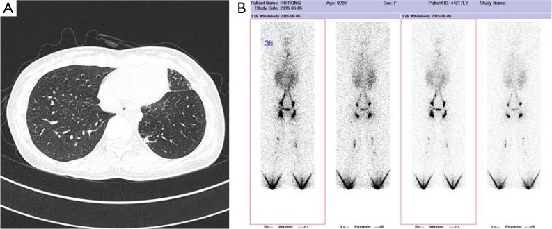
Figure 1.
A 28-year-old female with diffuse pulmonary lymphangiomatosis. (A) High resolution CT with diffuse pulmonary lymphangiomatosis. There is diffuse, smooth subpleural and interlobular septal thickening, peribronchovascular thickening is also present; (B) lymphangiography show lymphatic chain structure of both lower limbs is complete, and also lymphatic drainage can be unblocked.

