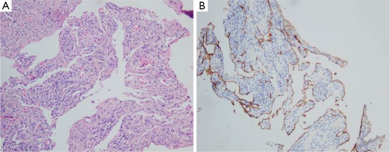
Figure 2.
A 28-year-old female with diffuse pulmonary lymphangiomatosis. (A) HE-section of lesion showing proliferation of thin-walled, anastomosing lymphatic vessels lined by single layer of endothelial cells lacking cytological atypia (arrows, 200×); (B) immunohistochemical staining with D2-40 revealing proliferative lymphatic channels (200×).

