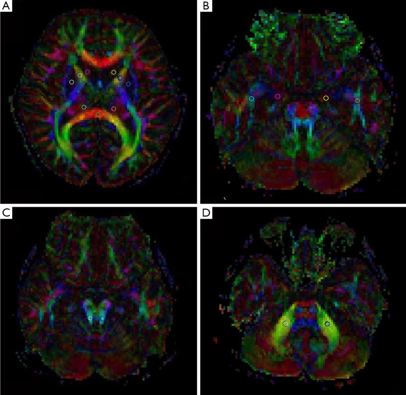
Figure 1.
Region of interest placement for DTI analysis. The circle indicates the location and size of the ROI. (A) Genu of corpus callosum, splenium of corpus callosum, caput nuclei caudati, lenticular nucleus, anterior limb of ICAL, posterior limb of ICPL, and Th; (B) temporal lobe WM and CgH; (C) SCP; (D) MCP. DTI, diffusion tensor imaging; ROI, outlined regions of interest; ICAL, internal capsule; ICPL, internal capsule; Th, thalamus; WM, white matter; CgH, cingulum in the hippocampus; SCP, superior cerebellar peduncle; MCP, diddle cerebellar peduncle.

