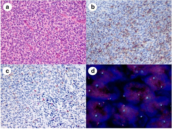
Fig 1.

Representative images of a malignant peripheral nerve sheath tumor mimicking a poorly differentiated synovial sarcoma. a: The tumor consisted of solid proliferations of uniform, round tumor cells with round to oval nuclei. b: Tumor cells were focally positive for S-100 protein on IHC. c: Tumor cells were sparsely positive for cytokeratin AE1/AE3 on IHC, d: FISH revealed no SS18 split signals
