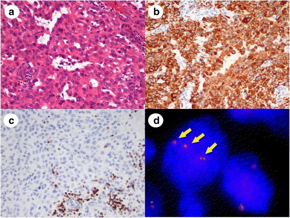
Fig 2.

Representative images of sarcomatoid carcinoma after synovial sarcoma was excluded. a: The tumor had a pseudoglandular structure and a sheet-like proliferation of cuboidal tumor cells. b: Tumor cells were positive for cytokeratin AE1/AE3 on IHC. c: Tumor cells were negative for bcl-2 on IHC. Infiltrating lymphocytes alone were bcl-2 positive. d: FISH analysis revealed no SS18 split signals. Nuclei showed more than two pairs of orange and green signals with a polyploidy pattern (arrows)
