Fig. 9.

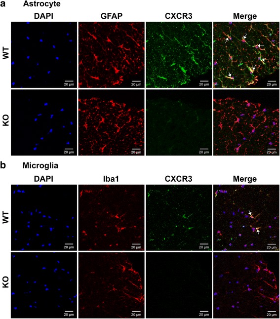
CXCR3 is expressed on both astrocytes and microglia. Frozen sections of lumbar spinal cord collected from WT and CXCR3−/− mice at the peak of disease (day 15) were subjected to immunofluorescence staining as described in the “Methods” section. a Double-labeling fluorescent staining shows CXCR3 on astrocytes. Anti-CXCR3 (green), anti-GFAP (red), DAPI (blue). b Double-labeling fluorescent staining shows CXCR3 on microglial. Anti-CXCR3 (green), anti-Iba1 (red), DAPI (blue). The arrows indicate co-localization of CXCR3 and glial cell markers. The immunofluorescence staining with isotype control antibody for anti-CXCR3 is shown in Additional file 5: Figure S5. Data are representative of three independent experiments
