Fig. 5.

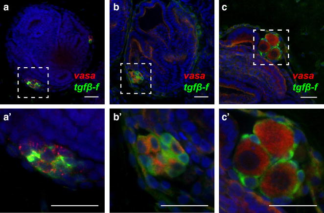
Organization of GFCs in oozooids, juveniles, and animals with maturing gonads. a–c, a′–c′) Confocal images of clusters composed of vasa-positive germ cells (red) and tgfβ-f-positive follicle progenitors (green) in Botryllus juveniles. The areas inside the dashed lines in a–c are displayed enlarged in a′–c′, respectively. a, a′ A GFC in a newly metamorphosed oozooid displays a lack of intracluster organization, with germ and follicle progenitor cells intermixed. b, b′ A GFC in an infertile juvenile also shows intermixed cell types and no intracluster organization. c, c′ Follicle cells envelop newly maturing stage 2 oocytes in a juvenile. Scale bars indicate 25 µm. Nuclei are shown by DAPI staining (blue)
