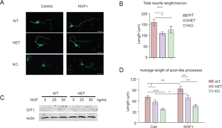
Figure 2.
Neurite growth was impaired in GIT1 knockout neurons.
(A) Hippocampal neurons isolated from wild type (WT), GIT1 heterozygote (HET) and GIT1 knockout (KO) mice at postnatal day (P) 0 were cultured for 5 days in control medium (left) or medium containing 50 ng/mL nerve growth factor (NGF) (right). Neurons were immunostained with anti-Tuj1 (green) antibody and DAPI (blue). Scale bar: 20 μm. Quantification of total neurite length per neuron (B) and average length of axon-like processes (D) of WT, HET and KO hippocampal neurons in vitro. The effects of NGF treatment are shown in Figure D. n = 30–50 cells; data are expressed as the mean ± SEM. The experiments were repeated three times. (C) Western blot of lysates from P0 WT and HET hippocampal neurons cultured for 5 days in the presence of 0, 25 or 50 ng/mL NGF. Anti-GIT1 and anti-actin antibodies were used to detect target protein expression. *P < 0.05, **P < 0.01, ***P < 0.001. One-way analysis of variance followed by Tukey's test.

